Tissue preparation cyropreservation.
Immunofluorescence protocol for frozen sections.
The suggested cryostat temperature is between 15 and 23 c.
Microscope slides pre coated.
Mount tissue sections onto gelatin or poly l lysine coated slides by placing the cold sections onto warm slides.
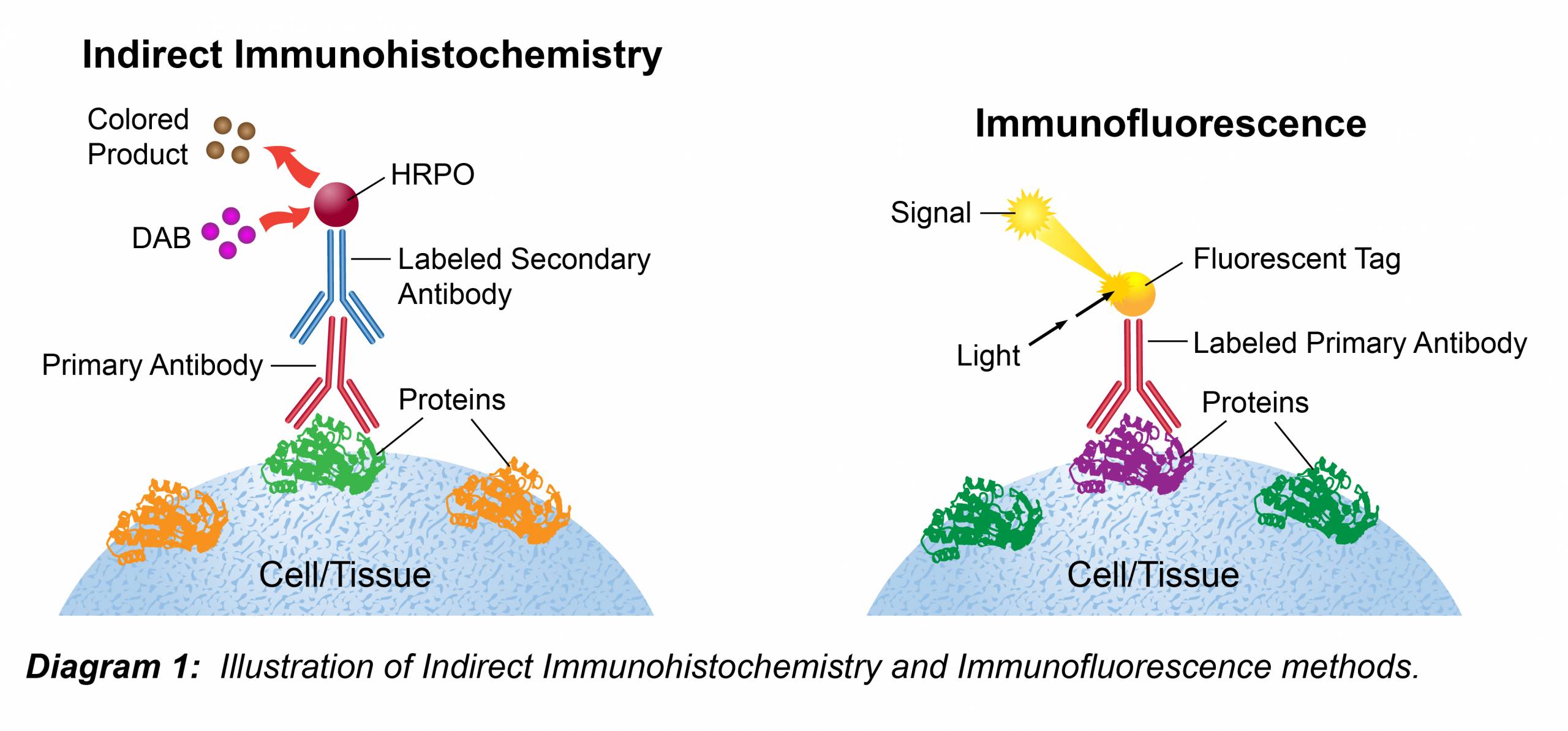
Immunofluorescence is commonly used to determine the cellular or tissue localization of a protein of interest.
Brigitte arduini version 1 2015 mar 23.
Nagy gertsenstein vintersten and behringer ed.
Store frozen blocks at 80 ºc.
Immunofluorescence staining protocol.
Materials phosphate buffered saline pbs 1x paraformaldehyde pfa 4 see support protocol 1.
Immunofluorescence on frozen sections.
Cryosections adhered to slides from blocks embedded in oct using the 2 methylbutane isobutene method.
Immunocytochemistry and immunofluorescence protocol related fluorescence.
The section will curl if the specimen is too cold.
Cut 4 8 um thick cryostat sections and mount on superfrost plus slides or.
Immunofluorescence can also be used as a qualitative measure of protein expression.
Please refer to the applications section on the front page of product datasheet or product webpage to determine if this product is validated and approved for use on cultured cell lines if ic paraffin embedded samples if p or frozen tissue sections if f.
Do not allow frozen tissue to thaw before cutting.
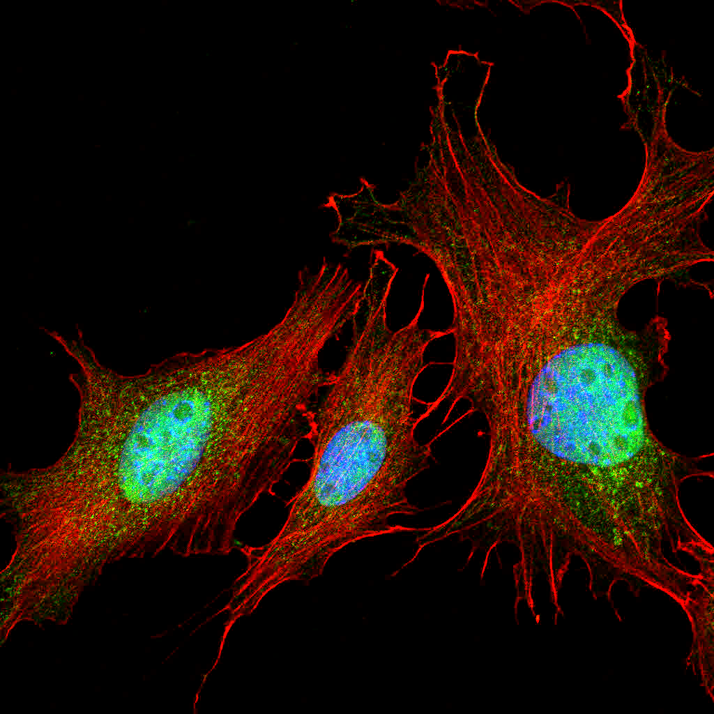
Annexin v labeled with alexa fluor 488 in frozen rat placenta section by ihc immunohistochemistry.
Cut cryostat sections at 5 10 µm and mount on gelatin coated histological slides.
Paraffin and frozen sections reagents can be applied manually by pipette or this protocol can be adapted for automated and semi automated systems if these are available.
Protocol for immunofluorescent staining of mouse frozen sections tissue.
Preparation of slides.
Modified from manipulating the mouse embryo 3.
Carry out incubations in a humidified chamber to avoid tissue drying out which will lead to non specific binding and high background staining.
This protocol is also suitable for 40µm free floating.
Icc and if video protocol.
The following is a general procedure guide for preparation and staining of acetone fixed frozen tissues using a purified unconjugated primary antibody biotinylated secondary antibody and streptavidin horseradish peroxidase sav hrp and dab detection system.
This portion of the protocol can be skipped if you are working with pre mounted tissue slides.
Immunohistochemistry protocol for frozen sections.
See cryoprotection and processing of embryonic tissue protocol.
Immunofluorescence general protocol important.
Snap frozen fresh tissues in liquid nitrogen or isopentane pre cooled in liquid nitrogen embedded in oct compound in cryomolds.
Slides can be safely stored for 6 12 months at 80 c until ready for staining.